The Visual Anatomy & Physiology Lab Manual offers a practical, visually oriented approach to understanding human anatomy and physiology through interactive exercises and modular organization․
1․1 Purpose of the Lab Manual
The Visual Anatomy & Physiology Lab Manual is designed to enhance student learning through a combination of visual aids, interactive exercises, and practical activities․ It serves as a comprehensive guide for a two-semester A&P lab course, providing students with hands-on experiences to explore anatomical structures and physiological processes․ The manual integrates with Mastering A&P, offering digital resources to reinforce concepts․ Its modular design ensures clarity and accessibility, making it an essential tool for active learning and academic success․
1․2 Structure and Organization
The Visual Anatomy & Physiology Lab Manual is structured into clear, two-page lab modules that focus on specific topics․ Each module combines visual aids, concise text, and practical exercises to enhance understanding․ The manual follows a body systems approach, aligning with typical A&P courses․ Its organization ensures logical progression, with visuals and text integrated seamlessly․ This design supports active learning and makes complex anatomical and physiological concepts accessible and engaging for students․
1․3 Key Features of the Manual
The manual features high-quality visuals, interactive simulations, and practical exercises․ It integrates with Mastering A&P, offering digital resources to enhance learning․ Two-page lab modules provide focused, self-contained lessons․ The combination of visual and hands-on activities promotes deep understanding of anatomy and physiology․ Safety protocols and post-dissection procedures are emphasized, ensuring a comprehensive lab experience․

The Visual Approach in Anatomy and Physiology
The visual approach combines high-quality images, videos, and interactive tools to enhance understanding of complex anatomical structures and physiological processes, making learning engaging and effective․
2․1 Importance of Visual Learning in A&P
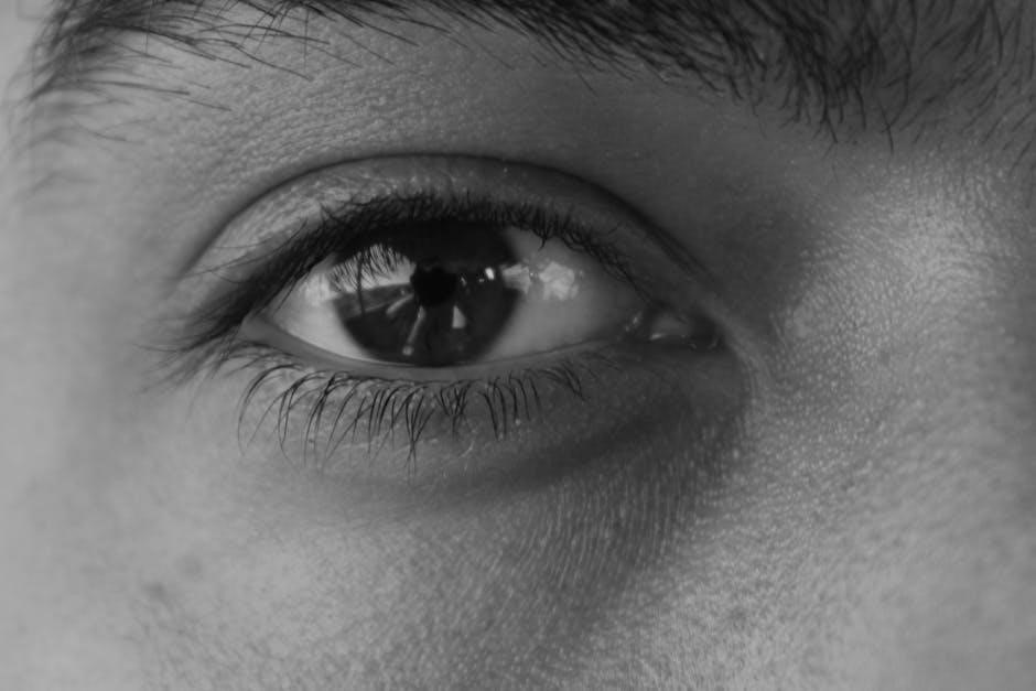
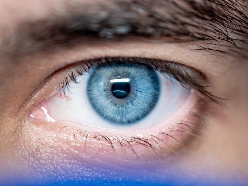
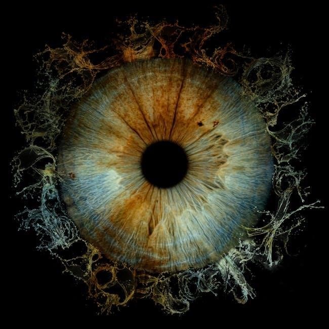
Visual learning is crucial in anatomy and physiology as it helps students grasp complex structures and processes more effectively; Images, diagrams, and videos complement textual information, enhancing memory retention and understanding․ Interactive tools and simulations further engage learners, making abstract concepts tangible․ This approach fosters a deeper connection with the material, improving overall academic performance and practical application in lab settings․
2․2 Using Images and Videos for Better Understanding
Images and videos are essential tools for enhancing comprehension in anatomy and physiology․ High-quality visuals provide clear representations of complex structures, while videos demonstrate dynamic processes like muscle movements or blood flow․ These resources allow students to engage with material in multiple ways, reinforcing learning and improving retention․ Interactive simulations and 3D models further enhance understanding, making abstract concepts more accessible and aiding in preparation for lab sessions and dissections․
2․3 Interactive Tools and Simulations
Interactive tools and simulations enhance learning by providing hands-on experiences with virtual models and real-time processes․ Students can explore 3D anatomical structures, simulate physiological functions, and conduct virtual dissections․ These tools foster engagement and deepen understanding through interactive exploration․ They also allow learners to visualize complex processes, such as nerve impulses or blood circulation, making abstract concepts more tangible and accessible for study and review․
Fundamentals of Anatomy and Physiology
This section explores the basic principles of anatomy and physiology, focusing on cellular structure, tissue types, and the organization of body systems, forming the foundation for advanced study․
3․1 Cellular Structure and Function
The cell is the basic structural and functional unit of life․ It consists of a cell membrane, cytoplasm, and nucleus, with organelles like mitochondria and ribosomes performing specialized functions․ Cells maintain homeostasis, reproduce, and respond to stimuli, forming the foundation of all biological processes․ Understanding cellular structure and function is essential for exploring anatomy and physiology, as it provides insight into how tissues, organs, and systems operate harmoniously․
3․2 Types of Tissues and Their Functions
Tissues are groups of specialized cells that perform specific functions․ Epithelial tissues form linings and glands, protecting surfaces and regulating exchange․ Connective tissues support, connect, and protect organs, with examples like bone and blood․ Muscle tissues enable movement through contraction, while nervous tissues transmit and process information․ Understanding these tissue types is fundamental for exploring how body systems interact and maintain overall health․
3․3 Organization of Body Systems
The human body is organized into interconnected systems, each specializing in specific functions․ The skeletal and muscular systems provide support and movement, while the nervous system controls communication․ The circulatory system transports nutrients, and the digestive system processes food for energy․ This modular organization allows for efficient functioning, with systems working together to maintain homeostasis․ Visual aids and practical exercises in the lab manual help students explore these relationships and understand their roles in overall physiology․

Laboratory Exercises and Activities
Laboratory exercises and activities are designed to engage students in hands-on learning, using visual tools and practical applications to explore anatomical structures and physiological processes effectively․
4․1 Planning and Preparing for Lab Sessions
Planning and preparing for lab sessions involves setting clear learning objectives, gathering necessary materials, and reviewing safety protocols․ Students should preview visual guides, familiarize themselves with equipment, and organize lab reports․ Proper preparation ensures efficient use of time, enhances engagement, and promotes a safe learning environment․ Utilizing digital resources and interactive tools beforehand can also deepen understanding of anatomical structures and physiological processes․
4․2 Safety Protocols in the Lab
Adhering to safety protocols is crucial in the lab to prevent accidents and ensure a secure environment․ Students must wear personal protective equipment (PPE) such as gloves and goggles․ Proper handling of sharp instruments and biological specimens is essential․ All chemicals should be used cautiously, and waste disposed of correctly․ Emergency procedures, like fire extinguisher locations, must be known․ Supervision and following instructions strictly are vital to maintain safety during experiments and dissections․
4․3 Conducting Dissections and Observations
Conducting dissections and observations requires precision and attention to detail․ The lab manual provides step-by-step guides to help students understand anatomical structures․ Practical exercises enhance observational skills, focusing on identifying key organs, tissues, and systems․ Students learn to use dissecting tools safely and effectively․ Observations are recorded for further analysis, fostering a deeper understanding of human anatomy․ This hands-on approach bridges theoretical knowledge with practical application, enriching the learning experience․
4․4 Data Collection and Analysis
Data collection and analysis are critical components of lab work․ Students learn to record accurate observations, measurements, and anatomical details during dissections․ Data is organized and interpreted to draw meaningful conclusions․ Practical exercises emphasize the importance of precision and scientific reasoning․ By analyzing collected data, students connect their findings to broader physiological concepts, enhancing their understanding of anatomical structures and their functions․ This process fosters critical thinking and scientific inquiry skills․
Visual Dissection Guides
Visual dissection guides provide step-by-step instructions and multimedia resources, such as images and videos, to enhance understanding of anatomical structures and their physiological functions․
5․1 Step-by-Step Dissection Procedures
The lab manual provides detailed, step-by-step dissection procedures, accompanied by high-quality images and videos․ Each procedure is divided into clear, manageable tasks, ensuring students can follow along effortlessly․ Pre-dissection preparation, anatomical identification, and post-dissection cleanup are all covered․ Multimedia resources, such as 3D models and interactive simulations, further enhance comprehension․ These guides are designed to promote active learning and engagement, making complex dissection processes accessible and engaging for students at all skill levels․
5․2 Identifying Key Anatomical Structures
The lab manual emphasizes the identification of key anatomical structures through detailed visuals and interactive tools․ High-quality images, 3D models, and real-time dissection videos guide students in recognizing and understanding complex structures․ Color-coded labels and clear descriptions aid in distinguishing between tissues, organs, and systems․ This visual approach ensures students can confidently identify structures, enhancing their ability to correlate anatomy with physiology in a practical and engaging manner․
5․3 Safety Measures During Dissection
The lab manual outlines essential safety measures to ensure a secure environment during dissections․ Students are required to wear protective gear, including gloves and goggles, to prevent exposure to biological materials․ Proper handling of sharp instruments and chemicals is emphasized, along with maintaining a clean workspace․ Clear guidelines for emergency procedures and waste disposal are provided to promote a safe and responsible learning experience․ Supervision by trained instructors is always recommended․
5․4 Post-Dissection Cleanup and Storage
Proper cleanup and storage are critical after dissection to maintain lab safety and organization․ Students are instructed to dispose of biological specimens and waste in designated containers․ Tools and equipment must be cleaned and stored in their assigned areas․ Workstations should be sanitized, and all materials must be labeled and securely stored for future use․ Following these steps ensures a tidy and safe learning environment for subsequent lab sessions․
Physiology of Major Body Systems
This section explores the structure and function of key body systems, such as muscular, nervous, and digestive, using visual aids to enhance understanding of physiological processes․
6․1 Muscular System: Structure and Function
The muscular system consists of skeletal, smooth, and cardiac muscles, enabling movement, support, and maintaining posture․ Visual aids like diagrams and videos illustrate muscle structure and contraction mechanisms, while exercises in the lab manual help students explore muscle physiology through dissection and observation, integrating with Pearson Anatomy and Physiology resources for a comprehensive understanding of muscle function and its role in overall body movement and stability․
6․2 Nervous System: Neural Communication
The nervous system facilitates neural communication through complex networks of neurons, synapses, and neurotransmitters․ Visual aids like detailed diagrams and 3D models illustrate how action potentials propagate and signals are transmitted․ The lab manual includes exercises to explore nerve structure and function, while interactive simulations, such as those in Mastering A&P, enhance understanding of synaptic transmission and the integration of neural pathways in controlling bodily functions and responses․
6․3 Digestive System: Nutrition and Absorption
The digestive system’s primary role is to break down food into nutrients for absorption and utilization by the body․ Through visual aids and interactive simulations, students explore the digestive tract’s structure and function․ Exercises focus on enzymatic processes, nutrient absorption in the small intestine, and the role of the liver and pancreas․ Mastering A&P supplements learning with 3D models and quizzes to reinforce key concepts of digestion, absorption, and the body’s nutritional needs․
Integration with Mastering A&P
The Visual Anatomy & Physiology Lab Manual seamlessly integrates with Mastering A&P, offering interactive simulations, quizzes, and digital resources to enhance learning and track student progress effectively․
7․1 Connecting the Lab Manual to Digital Resources
The Visual Anatomy & Physiology Lab Manual integrates seamlessly with Mastering A&P, providing access to interactive simulations, quizzes, and multimedia resources․ Students can use access codes to unlock digital tools, enhancing their learning experience․ The manual’s modular structure aligns with online materials, ensuring a cohesive approach to understanding complex concepts․ This integration supports active learning and allows instructors to track progress, making it easier to customize assignments and improve student outcomes effectively․
7․2 Using Interactive Simulations
Interactive simulations in Mastering A&P provide dynamic, hands-on learning experiences, allowing students to explore complex anatomical and physiological processes in detail․ These simulations include 3D models, virtual dissections, and interactive diagrams that enhance understanding of body systems․ Students can manipulate simulations to observe how structures and functions interact, reinforcing concepts covered in the lab manual․ This engaging approach makes abstract ideas more tangible and supports deeper comprehension of anatomy and physiology principles effectively․
7․3 Tracking Progress and Understanding
Mastering A&P provides tools to track student progress, offering insights into comprehension and areas needing improvement․ Interactive quizzes, assignments, and simulations generate performance metrics, enabling students to gauge their mastery of concepts․ Instructors can monitor progress through dashboards, identifying trends and supporting struggling learners․ This integration ensures a personalized learning experience, helping students achieve their goals effectively while aligning with the lab manual’s visual and practical approach to anatomy and physiology education․
Effective Learning Strategies
Effective learning strategies involve active engagement, visual aids, and practical exercises to enhance understanding and retention of anatomy and physiology concepts․
8․1 Active Learning in the Lab
Active learning in the lab involves hands-on activities, group discussions, and interactive exercises to engage students and enhance understanding of anatomy and physiology concepts․ By participating in practical tasks, students can visualize structures, conduct experiments, and apply theoretical knowledge․ This approach fosters critical thinking, collaboration, and problem-solving skills, making learning more effective and enjoyable․ Incorporating visual aids, simulations, and real-world applications further enriches the learning experience․
8․2 Practicing with Visual Aids
Visual aids like diagrams, videos, and 3D models are essential for mastering anatomy and physiology concepts․ By practicing with these tools, students can better understand complex structures and their functions․ Interactive simulations and labeled images allow learners to explore systems in detail, enhancing retention and comprehension․ Visual practice also supports active learning strategies, helping students connect theoretical knowledge with practical applications in the lab and real-world scenarios․
8․3 Making Connections Between Concepts
Making connections between concepts enhances understanding by linking anatomy and physiology to real-world applications․ Visual aids and interactive tools help integrate ideas, showing how systems interrelate․ By associating theoretical knowledge with practical observations, students can better grasp complex processes․ This holistic approach fosters deeper learning and prepares learners for clinical scenarios, emphasizing the importance of interconnectedness in human biology․

The Immune System: A Special Focus
The immune system is a vital component of human anatomy and physiology, protecting the body through specialized cells, tissues, and organs․ This section explores its mechanisms and interactions, essential for health and disease prevention․
9․1 Basic Concepts of Immunology
Immunology focuses on the immune system’s structure and function, emphasizing how it protects the body from pathogens and diseases․ Key concepts include innate and adaptive immunity, with cells like lymphocytes and macrophages playing central roles․ Antigens trigger immune responses, leading to antibody production and memory cell formation․ Understanding these processes is crucial for grasping how the body maintains health and combats infections․
9․2 Active Learning Strategies for Immune System
Engage with immune system concepts through interactive simulations, group discussions, and hands-on activities․ Use visual aids like diagrams and videos to explore how pathogens are recognized and neutralized․ Participate in case studies to analyze immune responses and their implications․ Apply critical thinking by predicting outcomes of immune system dysfunctions, fostering a deeper understanding of its role in health and disease․
9․3 Practical Applications in the Lab
Conduct hands-on lab exercises to explore immune system functions, such as observing blood smears to identify immune cells․ Use dissection guides to examine lymphoid organs like the spleen and lymph nodes․ Engage in simulations to model immune responses and experiment with virtual labs to understand vaccine development․ These practical activities bridge theory with real-world applications, enhancing understanding of the immune system’s structure and function․
Assessment and Evaluation
Evaluation includes quizzes, tests, lab reports, and presentations, ensuring comprehension of anatomy and physiology concepts through practical and theoretical assessments․
10․1 Quizzes and Tests
Quizzes and tests are essential tools for evaluating student understanding of anatomy and physiology concepts․ They assess comprehension of key topics and the ability to apply knowledge practically․
Regular quizzes ensure retention of detailed anatomical structures and physiological processes, while tests measure cumulative understanding․ Both formats use visual aids and case studies to reinforce learning and critical thinking skills;
10․2 Lab Reports and Presentations
Lab reports and presentations are crucial for assessing student understanding and communication skills․ Reports document experimental procedures, observations, and conclusions, fostering critical thinking and scientific writing․
Presentations enhance public speaking abilities, requiring students to clearly articulate complex anatomical and physiological concepts․ Both formats align with course objectives, ensuring comprehensive grasp of the material․
10․3 Dissection Practical Exams
Dissection practical exams assess students’ ability to identify and describe anatomical structures accurately․ These exams evaluate hands-on knowledge and understanding of spatial relationships between organs and tissues․
Students are required to demonstrate their skills in identifying key structures and explaining their functions․ Preparation involves thorough study of visual aids, dissection guides, and practice labs to ensure proficiency․
Be First to Comment